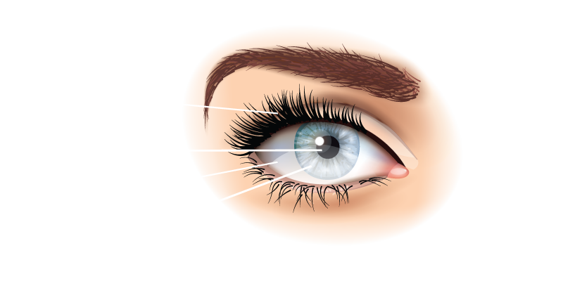
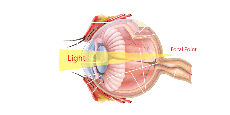
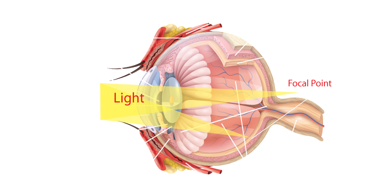
At the front of the eye is a transparent structure called the cornea. It is responsible for the majority of the focusing power of the eye.
Behind the cornea is the iris, a coloured tissue that defines someone’s eye colour, but it’s responsible for more than just the way a person appears. The iris expands or contracts, controlling the amount of light entering the eye.
Did you know that the iris is unique to each individual? More so than the corresponding fingerprints on each hand, as the iris is different between the two eyes too.
The iris forms a circular opening at its centre, known as the pupil.
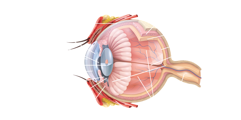
Between the cornea and the iris is an area called the anterior chamber, that is filled with clear fluid (the aqueous humour) which helps to provide nutrients inside the eye.
The white part of the eye is known as the sclera, which provides a tough protective coat to the layers inside the eye.
Attached to the sclera are the extraocular muscles which control the movement of the eyeball and its ability to change direction of gaze.
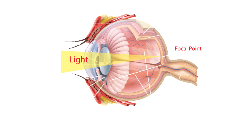
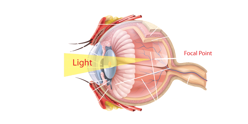
Behind the iris is located the crystalline lens. This lens is the only variable optical element in the eye, as it has the ability to change its shape via small muscles around the lens. This allows a person to change focus and see clearly both in the distance and up close. This process is referred to as accommodation. As we age, the lens material loses its elasticity, becomes harder, and the ability to focus on close objects will decrease.
After the light is focused by the cornea and the lens, it is transmitted to the retina. The retina covers almost the entire inner surface of the eye. It is made up of three layers of nerve cells, including light receptors. These cells are responsible for turning light into electrical signals which are then sent to the brain via the optic nerve.
In the centre of the retina is an area known as the macula, which is the most sensitive part of the human retina and is responsible for all fine detail vision.